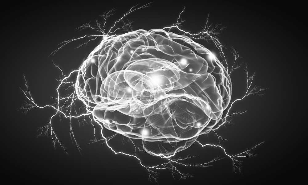
A recent article in Psychological Medicine reports that the grey matter changes seen in patients who undergo electroconvulsive therapy (ECT) do not account for the depression relief some patients reportedly experience post-ECT.
To the contrary, this study found that a post-ECT rise in grey matter volume corresponded with worsening depressive symptoms in the short run. This initial increase in grey matter volume was then succeeded by significant decreases over the subsequent two years. These reductions were correlated with deteriorating long-term outcomes. The authors state:
“Volume increases induced by ECT appear to be a transient phenomenon as volume strongly decreased 2 years after ECT. Short-term volume increases are associated with less symptom improvement, suggesting that the antidepressant effect of ECT is not due to volume changes. Larger volume decreases are associated with poorer long-term outcome, highlighting the interplay between disease progression and structural changes. “
The study was led by a multidisciplinary team, including Tiana Borgers, Verena Enneking, Melissa Klug, and many others. They are primarily affiliated with the Institute for Translational Psychiatry at the University of Münster in Germany. Additional contributors hail from institutions such as the University of Halle, the German Center of Mental Health in Halle, the University Hospital Frankfurt, and the University Hospital Jena. The collaboration also extended internationally with input from the Department of Psychiatry at the University of Melbourne and The Florey Institute of Neuroscience and Mental Health in Australia.
 The study aimed to analyze immediate and long-term changes in grey matter volume resulting from ECT and ascertain if such changes influenced clinical results. For this purpose, the authors enrolled 50 individuals with acute major depressive disorder (MDD) from the Department of Psychiatry at the University of Münster and the Landschaftsverband Westfalen-Lippe Hospital in Münster. Seventeen of these individuals underwent ECT, while 33 received medication and psychotherapy. The authors also enrolled 21 individuals without MDD using public advertisements and newspaper notices to serve as a control.
Individuals were excluded from participating if they had neurological issues, prior traumatic head injuries, organic mental conditions, chronic illnesses, current benzodiazepine use, a diagnosis of bipolar or psychotic disorders, a history of substance dependence, or health conditions preventing MRI scans.
All participants with MDD were undergoing a moderate to severe depressive episode and were receiving inpatient psychiatric care at the study’s commencement. Those in the ECT cohort were termed “treatment-resistant” as their symptoms hadn’t improved after trying at least two different antidepressant medications. Non-ECT MDD participants had never undergone ECT. The healthy control group had no history of psychiatric conditions and consistently scored below the threshold for depressive symptoms on all tests.
Before ECT, immediately after, and then two years post-ECT, the ECT group underwent MRI scans and two depression evaluations (the Hamilton Depression Rating Scale and the Beck Depression Inventory). Non-ECT groups were evaluated at analogous time intervals.
Both MDD groups noted substantial reductions in their depression test scores. 58.8% of ECT recipients saw at least a 50% score decrease, compared to 48.5% of the non-ECT MDD participants.
The ECT group experienced a notable grey matter volume spike immediately post-ECT, significantly decreasing two years later. The hippocampus, amygdala, inferior frontal gyrus, and insula were the most affected brain regions. Neither the non-ECT MDD group nor the healthy control group displayed significant grey matter volume shifts. However, minor reductions were noted in both groups’ 2-year MRI.
The ECT group’s increase in grey matter volume was tied to deteriorating depressive symptoms. Over the study’s 2-year duration, grey matter loss was also associated with poorer long-term outcomes for this group.
The researchers acknowledged several study limitations. The groups had small sample sizes, making broader implications challenging. A randomized control trial was not executed. The ECT group’s “treatment-resistant” nature might have affected its comparability to the non-ECT group. The authors didn’t explore potential symptom alleviation tied to grey matter volume changes in specific brain areas due to the need for more detailed scans. More frequent assessments might offer more accurate data on MDD progression.
They conclude:
“The association between GMV decreases and poor long-term outcome supports the transience of ECT-induced effects on structural and symptom level. Findings of notable GMV increases following ECT in areas that have been linked to the etiology and maintenance of MDD replicate immediate ECT-related effects found in previous studies. These GMV increases were associated with less immediate and delayed symptom improvement. Additionally, GMV changes were not found to be associated with changes in suicidality and delusion. Therefore, it remains elusive whether ECT-induced GMV increases are relevant for the antidepressant effect of ECT. The results seem to suggest that ECT-induced GMV increases are an epiphenomenon, while GMV decreases in the naturalistic long-term course of disease appear to be a correlate of relapse or ongoing depression, as stated by the neurotoxicity hypothesis.”
Studies have consistently found no discernible brain differences between those with and without depression. Presently, brain scans cannot distinguish between various mental health disorders. One research found disparate conclusions when multiple teams analyzed identical brain scan data.
Many experts deem findings from brain scan studies as “problematic if not unsubstantiated.” Some label MRI studies as inconsistent and unfit for research purposes.
A study in Nature pointed specifically to brain imaging research with small sample sizes as likely to produce unreliable conclusions.
****
Borgers, T., Enneking, V., Klug, M., Garbe, J., Meinert, H., Wulle, M., König, P., Zwiky, E., Herrmann, R., Selle, J., Dohm, K., Kraus, A., Grotegerd, D., Repple, J., Opel, N., Leehr, E. J., Gruber, M., Goltermann, J., Meinert, S., Bauer, J., … Redlich, R. (2023). Long-term effects of electroconvulsive therapy on brain structure in major depression. Psychological medicine, 1–11. Advance online publication. https://doi.org/10.1017/S0033291723002647 (Link)


“ECT” = ELECTRO-CUTION TORTURE
Richard Sears does a good job of presenting the WORD SALAD of psychiatry in an appetizing way. He is not the target of my criticism. Psychiatry itself, and the genocidal practice of “ECT” is my target. How can anybody think that a non-fatal electrocution is “health care”? The idea is absurd on the face of it. I fail to understand, how in 2023, people with actual college degrees can get paid fat 5 & 6 figure salaries, and still believe the “ECT” bullshit. Oh, I guess I answered my own question. The psychs torture people for MONEY. Makes sense now….
“The love of money is the root of all evil.”….
Sometimes, the Bible DOES get it right….
Report comment
Ever since psychiatrists introduced the new “modern” ECT machines in the 1980’s that are 4-6x stronger than previous sine machines initially used, they have refrained from completing any legitimate neuropathology studies on ECT recepients. They point to a single study of a poor, incontinent, speechless patient who had more than 1000 ECT treatments across the course of their lifetime as proof this patient had no damage. Reading their “proof” reveals they did not complete a modern neuropathology study with proper staining on that person.
Instead they seemed to have removed the brain and examined it visually–no staining, no microscope, no slides in the “study” to prove the brain was as healthy as they claimed.
Even the football players brains’ look “normal” without proper staining under a microscope.
There’s a reason Psychiatrists have chosen not to conduct modern neuropathology tests on ECT recepients. It may have everything to do with major gaps in their own medical training and cognitive dissonance–a stubborn refusal to recognize repeatedly spiking cerebral profusion 300% and glucose and oxygen consumption 200% has lasting, progressive consequences just like any other form of repetitive mild-moderate traumatic brain injury and repetitive electrical injury.
Report comment
Not to mention profits!
Report comment